Applications
Material analysis.
Fibres, pores and particles.
Inspection tasks
Structure, measured in three dimensions.
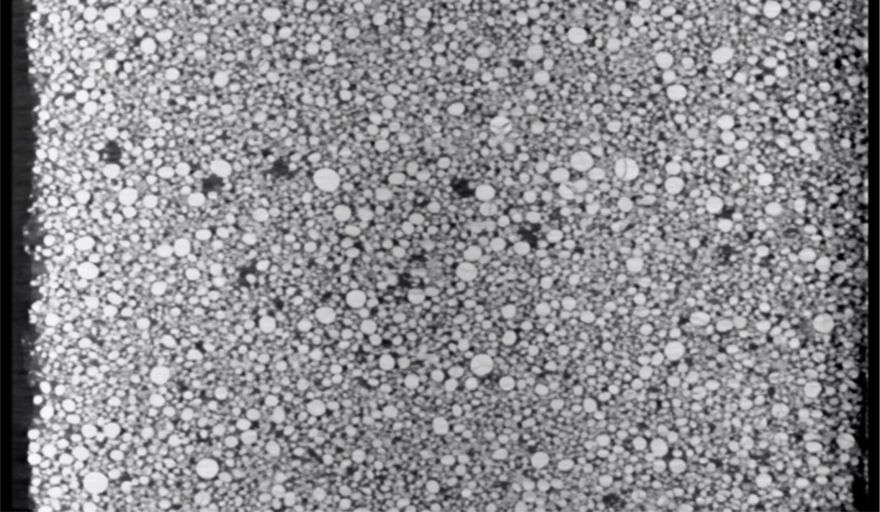
Pores, fibres, phases and particles determine how a material behaves. High-resolution CT quantifies them in the volume and leaves the specimen intact for the next test.
Porosity and phases
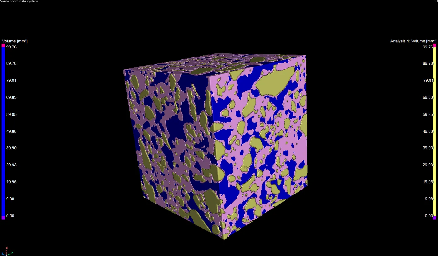
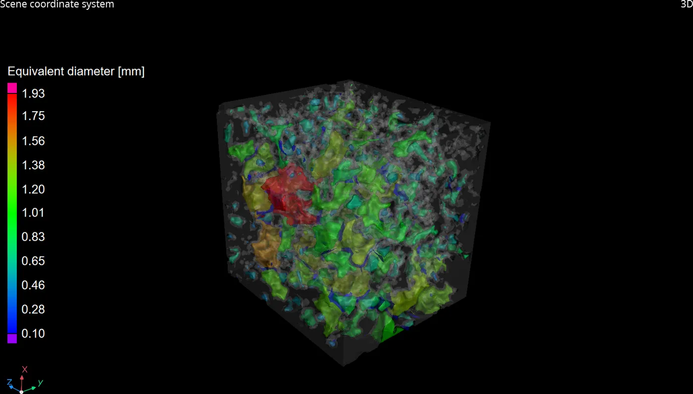
Pore size distribution and phase fractions are calculated from the volume, not estimated from a section.
Composites and ceramics
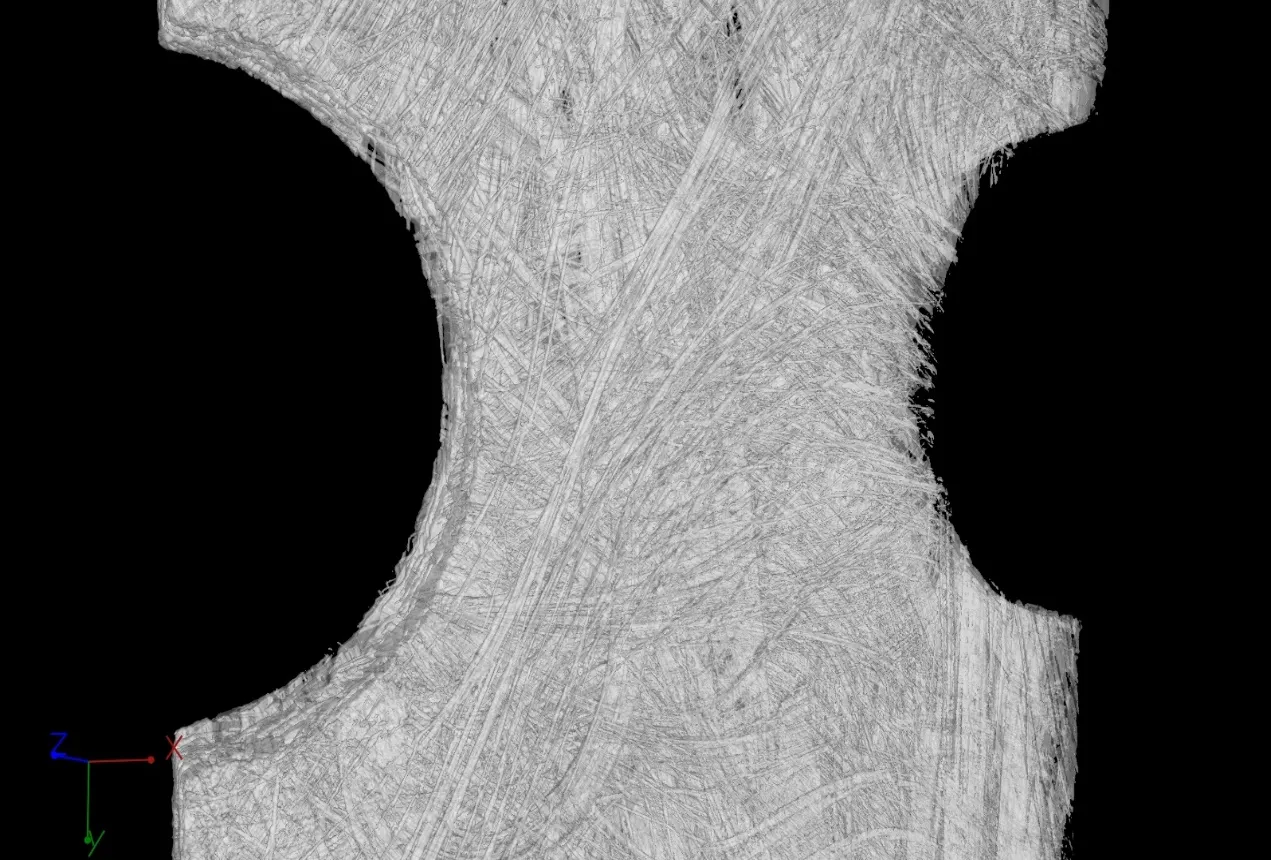
Fibre structure, delamination and micro-cracks are shown in composite and ceramic materials.
Rock, concrete and powders
Geotechnical specimens, concrete and particle agglomerates are analysed without preparation.
All these inspection tasks are covered in detail in our material analysis presentation.
Scan examples
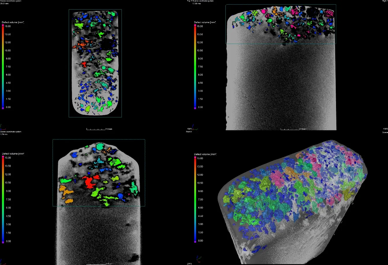
What the data shows.
Systems
Quantified structure, intact specimen.
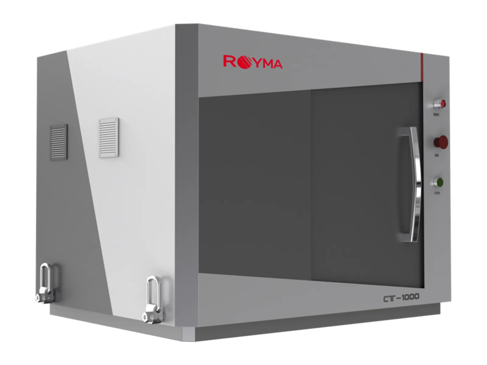
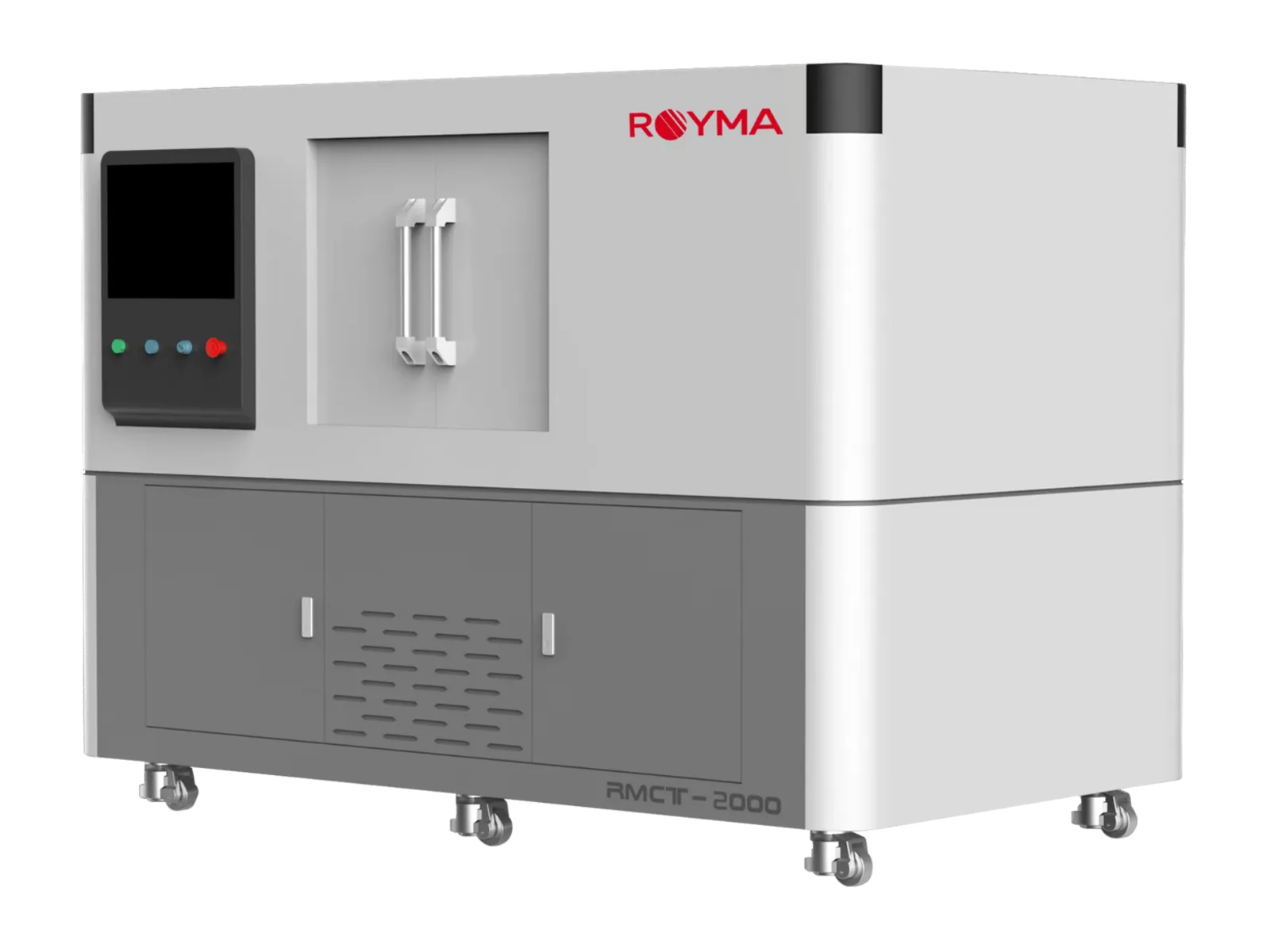
Royma offers high-resolution CT for materials research and materials testing. The RMCT2000 resolves the structure of a specimen at 0.9 µm spatial resolution or better in its 190 kV nanofocus mode and switches to a 225 kV microfocus mode for larger samples up to ø 300 × 250 mm. The desktop RMCT1000 gives a laboratory a 4 µm CT on the bench, with six scan modes and one-click imaging for routine specimens.
The volume is the measurement: pore size distribution, phase fractions, fibre orientation, particle size and the geometry of cracks and delaminations are calculated from the three-dimensional data set, not estimated from a polished section. Research groups and test laboratories gain a non-destructive method that leaves the specimen available for the mechanical test that follows.
Composites, ceramics, foams, rock, concrete and powder agglomerates are scanned without preparation, and the same specimen can be scanned again after loading or ageing to follow the change.
In practice
From scan to statistics.
The analysis software segments the phases and pores in the volume and returns the distributions as tables and histograms, together with the rendered volume for inspection. Fibre orientation tensors, strut thickness in foams and crack networks are evaluated with the same tools.
Scan recipes are stored per specimen type, so a series of specimens is evaluated identically and results are comparable across a study.
Beyond the laboratory bench, the RMCT4000 and RMCT6000 extend the same evaluation to larger specimens and dense materials: cores, cast test bars, welded coupons and structural samples are scanned with microfocus and mini-focus sources up to 600 kV, and the porosity or crack evaluation runs with the same software as on the small specimen.
Questions
Questions about CT for material analysis.
- What resolution is available for materials specimens?
- The RMCT2000 reaches a spatial resolution of 0.9 µm or better in nanofocus mode; the RMCT1000 offers 4 µm or better on the bench.
- Can porosity and phase fractions be quantified?
- Yes. The volume is segmented into phases and pores, and the software returns pore size distribution, phase fractions and particle statistics for the whole specimen.
- Is the specimen still usable after the scan?
- Yes. CT is non-destructive; the specimen remains intact for mechanical testing or for a second scan after loading or ageing.
- Which materials can be scanned?
- Metals, polymers, composites, ceramics, foams, rock, concrete and powders; the sample size and the density decide between nanofocus and microfocus mode.