
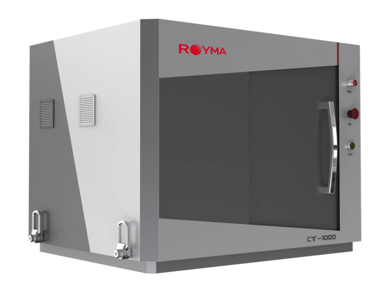
- Closed X-ray tube, 40 W
- 130 kV
- Spatial resolution
- ≤ 4 µm
- Maximum scannable sample size
- ø 210 × 140 mm
- System weight
- 0.9 t
Overview
CT on a laboratory bench.
Not every laboratory has room, or budget, for a CT cabinet. The RMCT1000 is a desktop system that brings micrometre resolution to small samples.
One-click imaging runs the scan from start to volume. Occasional users get results without CT training.
It is made for small, light samples. Dense metal parts and anything above ø 210 × 140 mm belong in a cabinet system.
Features
Small system. Full CT.
Resolve fine detail
Inspect small parts at micrometre level. The 130 kV closed tube and a detector with 49.5 µm pixels give a spatial resolution of 4 µm or better.
Start with one click
Get from sample to volume without set-up work. One-click imaging runs acquisition and reconstruction in one sequence.
Six scan modes
Use the scan that fits the sample. Circular, helical, segmented, offset, limited-angle and jitter scans are available.
Fits the room
Place it where the work is. The system measures 1357 × 820 × 900 mm, weighs 0.9 t and keeps radiation leakage below 0.5 µSv/h.
Specifications
Technical data.
| X-ray source | |
|---|---|
| Type | Closed tube |
| Maximum tube voltage | 130 kV |
| Maximum power | 40 W |
| Image quality | |
| Spatial resolution | ≤ 4 µm |
| Detector | |
| Effective area | 114 × 146 mm |
| Pixel size | 49.5 µm |
| Pixel matrix | 2304 × 2940 |
| Sample | |
| Maximum scannable size | ø 210 × 140 mm |
| Chamber size | ø 480 × 370 mm |
| Scan modes | |
| Available | Circular, helical, segmented, offset, limited-angle, jitter |
| Mechanics | |
| Axes | 5 |
| Installation | |
| System (L × W × H) | 1357 × 820 × 900 mm |
| Weight | 0.9 t |
| Radiation leakage | < 0.5 µSv/h |
Applications
Where it is used.
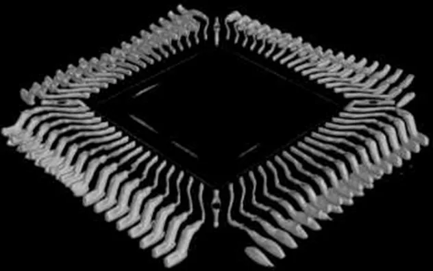
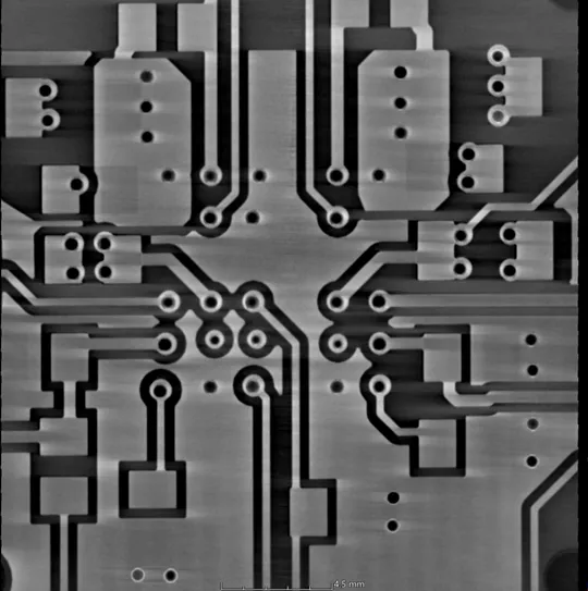
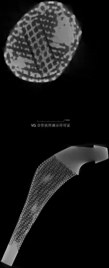
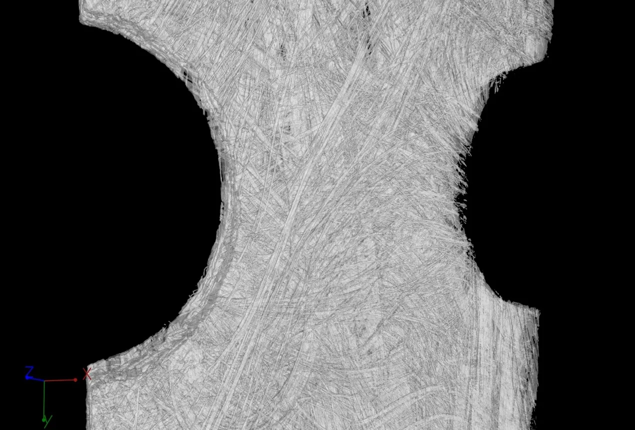
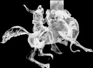
Pores, cracks, inclusions and assembly faults in small components, material samples and research objects.
Downloads
Data sheet on request.
We send the current RMCT1000 data sheet together with a first assessment of your part.