
- Linear accelerator source, six energy options
- 1–9 MeV
- CT spatial resolution, better than
- 2.5 lp/mm
- Maximum focus–detector distance
- 4000 mm
- Maximum sample load
- 1000 kg
Overview
When X-ray tubes stop, it continues.
Penetration is the hard limit of tube-based computed tomography (CT). The RMCT10000 replaces the tube with a linear accelerator. It inspects large castings, thick welds and assembled battery modules that otherwise have to be cut open.
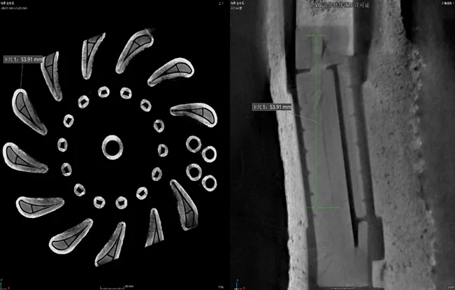
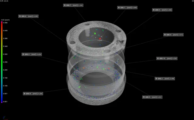
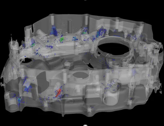
You get volume data of the complete component: porosity, shrinkage cavities, cracks, inclusions and weld defects, located in three dimensions.
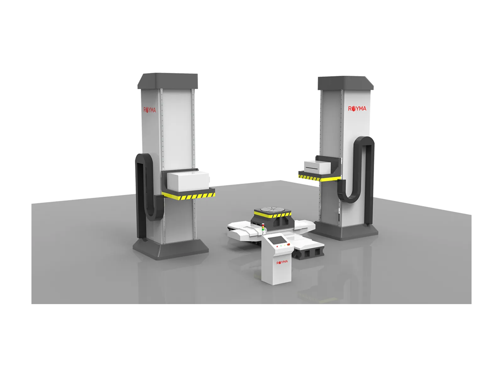
The RMCT10000 is a room installation, not a cabinet. For parts that a conventional X-ray tube penetrates, a Royma cabinet system is the simpler choice.
Features
Built for the thickest parts.
Penetrate dense material
Match the source to the part. The linear accelerator is available with 1, 2, 3, 4, 6 or 9 MeV electron energy.
Keep fine detail
See small defects in thick sections. A 1.5 mm focal spot supports a CT spatial resolution better than 2.5 lp/mm and a density resolution better than 0.5%.
Two detectors, one system
Choose speed or contrast. The flat panel covers 556 × 635 mm with 100 µm or 139 µm pixels. The 16-bit line detector rejects scatter with a post-collimator adjustable from 0.15 to 5 mm.
Scan modes for every shape
Fit tall, wide and awkward parts. Cone-beam CT runs as circular, offset, helical or segmented scan; the line detector adds fan-beam and offset fan-beam scans.
Whole components, not coupons
Scan the part as it is built. A stitched cone-beam scan covers ø 720 × 2600 mm, a fan-beam scan ø 1250 × 2300 mm, with a sample load of up to 1000 kg on the manipulator.
Planned into your building
The system is laid out for your hall. The standard room measures 6000 × 2500 × 3000 mm and can be adapted, with full X-ray protection and a safety interlock.
Specifications
Technical data.
| X-ray source | |
|---|---|
| Type | Linear accelerator |
| Electron energy | 1 / 2 / 3 / 4 / 6 / 9 MeV, by configuration |
| Focal spot | 1.5 mm |
| Dose rate | Up to 4500 cGy/min at 1 m with the 9 MeV source |
| Collimator angle | 0°–20° vertical and horizontal, motorised |
| Leakage outside the main beam | ≤ 0.1% of the central-axis dose rate, at 1 m from the target |
| Detectors | |
| Flat panel pixel size | 100 µm or 139 µm, selectable |
| Flat panel active area | 556 × 635 mm |
| Line detector pixel size | 0.2 mm or 0.4 mm |
| Line detector length | 614 mm or 820 mm |
| Line detector post-collimator | 0.15–5 mm, adjustable |
| Scan modes | |
| Cone beam | Circular, offset, helical, segmented |
| Line detector | Fan beam, offset fan beam |
| Image quality | |
| CT spatial resolution | Better than 2.5 lp/mm |
| CT density resolution | Better than 0.5% |
| Radiography, IQI sensitivity | Better than 1% |
| Sample | |
| Cone-beam scan, stitched | ø 720 × 2600 mm |
| Fan-beam scan, offset | ø 1250 × 2300 mm |
| Placeable sample on the manipulator | 2700 × 1400 mm |
| Load capacity | Up to 1000 kg |
| Mechanics | |
| Maximum focus–detector distance | 4000 mm |
| Controlled axes | Up to 6 |
| Main frame | Granite or cast iron |
| Installation | |
| Standard room | 6000 × 2500 × 3000 mm, customisable |
| Radiation protection | Full X-ray protection room with safety interlock |
Applications
Where it is used.
Porosity, voids, shrinkage cavities, cracks, inclusions and weld defects in very thick or dense components.
Downloads
Data sheet on request.
We send the current RMCT10000 data sheet together with a first assessment of your part.